A simple
chest X-ray (also called a roentgenogram, after Wilhelm Röentgen,
who discovered the X-ray in 1895) is often the first
imaging done of the heart. It is also the most commonly performed
imaging procedure. According to the American Journal of Roentgenology,
chest X-rays are taken in almost 20% of all Emergency Room visits:
22 million were done in 2003.
The X-ray
exposure
is minimal to the patient -- according to the American Heart
Association, it's about 1/5 of the annual exposure one normally
gets from
natural sources,
such as the
sun.
Frontal and
lateral chest X-ray views
made with T.RAD Plus Digital
system**
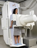
Standalone
X-ray unit**
Chest X-rays show
the size, shape and position of the heart, lungs and bone structures
of the chest. Sometimes your physician may order two views: a frontal
and lateral (side) view.
A chest X-ray can
be made at the patient's bedside, or in a
dedicated
room. The
latest
digital units eliminate film entirely and
allow for wider coverage at low radiation
levels.
The chest X-ray can tell your physician if your
heart has defects, is enlarged, has significant calcification,
pulmonary blood flow or if there is fluid in the lungs, sometimes
the result
of
a heart attack. The chest X-ray is
a preliminary procedure and is sometimes not necessary if the physician
has ruled out congestive heart failure and heart defects, since
the X-ray shows only the exterior shapes of the heart and surrounding
areas.
It
does not image the interior chambers or arteries.
Who Does
the Procedure: Chest X-rays are
usually done by a Radiology Technologist (RT) and interpreted
by a Radiologist.
Patient
Preparation:No preparation
is necessary before getting a chest X-ray, other than removing
metal objects such as necklaces and other metallic objects. etc.
It is very important to let the technologist
know
if you are
pregnant:
X-rays
are
not normally done on women who are pregnant. The
amount of radiation exposure is small for an adult, but can affect
a developing fetus.
send comments & suggestions
to "info at angioplasty dot org"
Read our Privacy statement.
Angioplasty.Org is an editorially independent informational health
site
which has received unrestricted educational grants from Medtronic plc,
TCROSS NEWS, Toshiba
America
Medical Systems, Volcano
Corporation, Terumo
Medical Corporation
Cardium Therapeutics, Inc. and Lenox Hill Heart and Vascular Institute of NY