Magnetic Resonance Imaging
Imaging and Diagnosis Section is Supported by TOSHIBA America Medical Systems
PatientCenter
VascularTherapy.OrgBlogDiscussion Forum
Home » Imaging & Diagnosis » Magnetic Resonance

Magnetic resonance imaging (MRI) or magnetic resonance angiography (MRA) are non-invasive methods of imaging internal organs, using radiofrequency waves and magnetic fields -- no X-ray radiation is involved and no iodine-containing contrast is used -- especially important for patients who have renal (kidney) disease and who may be negatively affected by the contrast used in standard or CT angiography.

MRI and MRA have limitations, however, in that highly detailed views of the coronary arteries may be better imaged using MultiSlice CT angiography or cardiac catheterization.

    
Ultra "open" MRI system
Ultra "Open" MRI imaging system**

Vantage wide-bore MRI system
Vantage wide-bore MRI imaging system**
   

While the use of MRI scans in cardiac applications requires an operator specifically trained in these uses, the technology is rapidly evolving and offers many benefits with minimal impact on the patient. Many experts in the field of cardiac imaging feel that MRI technology will become the primary tool for cardiologists in the future.

The original MRI units required the patient to lie still inside of a narrow "closed" environment. Due to the claustrophobic nature of this test and discomfort of patients, manufacturers designed an "open" system as well.

The test takes about 30-45 minutes and, since there is no radiation, a family member or friend may be in the room with the patient.


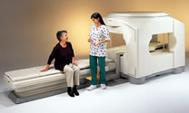
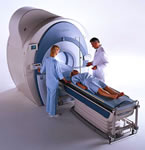
MRI or MRA can provide significant information about the presence or absence of coronary artery disease with very minimal impact on the patient and no radiation exposure, although it may not image the coronary arteries as precisely as the equally non-invasive MSCT angiogram.


Who Does the Procedure: MRI or MRA procedures are evaluated similarly to the MSCT, by a radiologist or cardiologist trained in this modality. A technologist usually performs the actual procedure.


Patient Preparation: Since MRI affect metals, if you have had any metallic implants (pacemakers, stents, surgical pins, etc.) or any metal in your body, you should let your physician know (although stents have been cleared for MRI immediately after placement). If you are pregnant, let the technologist know.


Electrocardiogram (EKG / ECG)

Chest X-ray

Exercise Stress Test with
Echocardiography (Ultrasound)

Nuclear Stress Test (Thallium or Technetium)

MultiSlice Computed Tomography and
CT Angiogram (CTA)

Magnetic Resonance Imaging and Angiography (MRI and MRA)

Cardiac Catheterization

Intravascular Ultrasound (IVUS)


** photo courtesy of Toshiba America Medical Systems

Angioplasty.Org Home •  PatientCenter


send comments & suggestions to "info at angioplasty dot org"
Read our Privacy statement.

Angioplasty.Org is an editorially independent informational health site
which has received unrestricted educational grants from
Medtronic plc, TCROSS NEWS, Toshiba America Medical Systems, Volcano Corporation, Terumo Medical Corporation
Cardium Therapeutics, Inc. and Lenox Hill Heart and Vascular Institute of NY

For more information, see About Us
All content, including text, photos and video
©Copyright Venture Digital LLC 1996-2021