Exercise Stress Test
Imaging and Diagnosis Section is Supported by TOSHIBA America Medical Systems
PatientCenter
VascularTherapy.OrgBlogDiscussion Forum
Home » Imaging & Diagnosis » Exercise Stress Test

An EKG, chest X-ray or other measurements, such as blood pressure, cholesterol tests, etc., are the first steps to diagnosing any cardiac problems. They are standard, routine, non-invasive and can yield information to guide you and your physician to the next steps.

If the patient has continuing symptoms, or if the above tests reveal any abnormalities that might be heart-related, the next diagnostic 
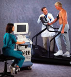
procedure is normally an exercise stress test, which is usually done 
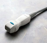
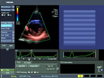
with "echo", short for "echocardiography", a non-invasive imaging of the heart that uses ultrasound. Ultrasound uses sound waves beyond the range of human hearing to image the chambers and walls of the heart. There is no radiation and the test is non-invasive.

In some cases physicians recommend going directly to a Thallium Exercise Stress Test and bypassing the Echo Exercise Stress Test. An exercise stress test with a Thallium Scan is more expensive and time-consuming than a stress test with ultrasound, but may reveal more information. Patients should discuss with their doctors what testing sequence is recommended.

An exercise stress test with echo allows the physician to see how the patient's heart is functioning while at work. A stress test may not be indicated for certain patients with known heart disease or other conditions. Some patients who may not be able to exercise using the machines can still take the test. A drug or pharmacologic agent, such as adenosine or dobutamine, can be administered to simulate the rapid heart beat achieved during exercise.


First part of the test is a "rest echo" or baseline study.

The patient is hooked up to an EKG and blood pressure cuff to help synchronize the study and to monitor the heart and blood pressure during the stress part of the test. A technician then glides a small hand-held ultrasound transducer around the patient's chest (a little clear gel is spread on the chest to ensure good contact).

The moving images are transmitted to a monitor and the cardiologist interprets them to see how efficiently your heart is pumping (the ejection fraction), thickness of the muscle walls, or if there are any valve leaks.

    
ultrasound transducer
typical transducer used in echocardiography**

treadmill test
Aplio CV ultrasoundimaging with treadmill**
   

When the resting study is completed, the exercise stress part begins. Placing the heart in a situation where it needs to pump blood more rapidly may reveal any blockages or other problems. For example, your car may sound fine on idle, but when you stress the engine at highway speeds, timing problems will become more evident.

The exercise is usually done using a treadmill, with variable speed and slant, but sometimes a stationary bicycle is used, often with the patient on his or her back to facilitate using the echo. The cardiologist will put the patient through the paces, going from warm-up to a level just short of the normal target rate for the patient's age and condition. The patient is monitored constantly and, if any EKG changes, chest pain or other concerning symptoms occur, the test is stopped. While this type of distress is rare, the cardiologist is always with the patient and can quickly provide relief.


When the stress test has reached its target, the echo study is repeated. All images are recorded to tape or disk, and the cardiologist can then analyze the rest and stress studies to determine if there are any problems.

Modern cardiac ultrasound systems can provide significant assistance in these analyses -- using color and Doppler studies to help the cardiologist interpret the study.

The main fact that can be learned is whether or not the heart acts normally when stressed. If any abnormalities show up, further testing to determine the cause is indicated.

    
Aplio echo doppler study
Aplio Tissue Doppler Imaging**

While not foolproof, an accurate stress echo test is able to diagnose heart disease or rule it out 85-90% of the time. Even if the test is normal, if the patient's symptoms persist, the cardiologist may schedule more testing.

Who Does the Procedure: An exercise stress test with echocardiography is normally performed by a cardiologist and a technician or nurse. It can be performed in a doctor's office, medical center or hospital.


Patient Preparation: You should not eat or drink anything for three hours before the test, since you will be exercising up to your capacity and this may cause nausea. If you are diabetic or are taking heart medications, your doctor will give you specific instructions on what to bring or what to stop taking before the test. Wear comfortable clothing since you will be "running a race".


Electrocardiogram (EKG / ECG)

Chest X-ray

Exercise Stress Test with
Echocardiography (Ultrasound)

Nuclear Stress Test (Thallium or Technetium)

MultiSlice Computed Tomography and
CT Angiogram (CTA)

Magnetic Resonance Imaging and Angiography (MRI and MRA)

Cardiac Catheterization

Intravascular Ultrasound (IVUS)


** photo courtesy of Toshiba America Medical Systems

Angioplasty.Org Home •  PatientCenter


send comments & suggestions to "info at angioplasty dot org"
Read our Privacy statement.

Angioplasty.Org is an editorially independent informational health site
which has received unrestricted educational grants from
Medtronic plc, TCROSS NEWS, Toshiba America Medical Systems, Volcano Corporation, Terumo Medical Corporation
Cardium Therapeutics, Inc. and Lenox Hill Heart and Vascular Institute of NY

For more information, see About Us
All content, including text, photos and video
©Copyright Venture Digital LLC 1996-2021