Whether it's called
multislice CT (MSCT), multidetector CT (MDCT), cardiac CT or cardiovascular
CT, the CT stands for "Computed
Tomography", a way of measuring parts of the anatomy by sections
(originally known as "Computed Axial Tomography" or CAT scans).
Single CT scans are widely used in medical imaging, but have been
less useful for imaging the heart, since a beating heart doesn't
stand still for a picture.
Now the development of multiple detectors,
or Multislice CT systems has allowed imaging of the heart, which
moves as it beats, with a level of detail not previously available.
If EKG's, stress tests or other
indicators have revealed a potential cardiac problem, the next
step is for the cardiologist to get a close-up look at the arteries
in the heart, to see where there might be blockages.
A few
years ago the patient would move on to a cardiac
catheterization,
during which a physician inserts a
catheter into the circulatory system, advances it to the heart
and injects dye into the coronary arteries. The physician then
makes an X-ray (cine-angiography)
movie from several different
angles, develops the film and sits at a viewing machine to analyze
the 35mm motion pictures. More recently, the film may have been
replaced by digital video. In either case, because the X-ray is
a "shadow" image that is two-dimensional, the cardiologist
has to interpret a number of different "camera angles" that
were shot to determine the presence and shape of any obstructions
to
the blood flow.
But a
much better diagnostic option would be a detailed 3D virtual model
of the patient's heart --
so that the cardiologist
could rotate, zoom and move through the heart's anatomy at any
angle at will, as if it were a video game. And without impacting
on the patient!
Multislice CT angiography provides
this 3D model, and it is revolutionizing cardiac imaging to the
point where a separate Society
of Cardiovascular Computed Tomography (scct.org) has recently
been formed to set standards for training and interpretation.
Multislice CT systems with 16 or
more detectors have made a quantum leap into imaging of the coronary
arteries.
A 256-slice system is now in development that will image the
entire heart in a single beat.
In less than 30 minutes,
without the invasiveness of a cardiac catheterization, a patient
can have an MSCT done to determine if there are any arterial blockages
that require an intervention, such as an angioplasty or stent.
The only immediate impact on the patient is an IV with iodine-containing
contrast and possibly a beta-blocker to
slow the heart beat (the
equivalent of saying "cheese" and holding still for the photographer).
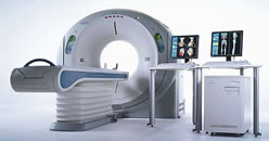
This is an actual 3D Multislice CT
scan made on an Aquilion™ 64-slice
CT scanner**
Aquilion™ 64-slice
CT scanner**
Although
MSCT does entail a higher radiation dose from that experienced
during a standard catheterization, mitigating
this risk is the fact that the patient is not exposed
to the complications that sometimes accompany cardiac catheterization
(angiography). Additionally, newer technology developments and
scanning methods that reduce the necessary radiation exposure are
ongoing.
If no blockages are found, MSCT provides a less
invasive and less
expensive method of ruling out the need for additional intervention.
If significant blockages
are found, then the patient is referred to a cardiac catheterization
with a probable angioplasty or stent. If previous tests show
a very high
likelihood
that the patient has significant coronary artery disease, then
MultiSlice CT angiography probably is not indicated, because the
patient will no doubt have to go to interventional treatment anyway.
An interesting feature of MSCT scans comes into play
if some disease is found, but it is not advanced enough to require
revascularization
using angioplasty or stenting. Physicians have reported
that when patients see such a clear and understandable picture
of their heart, they are much more motivated to make lifestyle
and other changes to lower their risk factors.
As MultiSlice CT becomes more widespread, it is likely that several
of the tests described in this section will become less and less
used. Some cardiologists think that nuclear stress testing will
be replaced by MSCT. For certain patients, MSCT will also replace
the "gold standard" of cardiac catheterization.
This is a rapidly developing technology. Methods of characterizing
the type of plaque in the arteries are being refined, so that
"vulnerable" plaques that are more likely to rupture can be pinpointed
for treatment, reducing the risk of heart attack. Right now,
MSCT provides a significant "next step" to
patients whose stress tests have proven inconclusive, and who may
have a good chance of being "screened out" for coronary
artery disease.
Who Does
the Procedure: MSCT can be done
by a cardiologist, radiologist or technologist trained in this
specific imaging modality. Because the MSCT scan reads the entire
upper
torso,
a growing number of heart and vascular centers are having both
cardiology and radiology specialists"read"
the results
-- the
cardiologist
for coronary disease, and the radiologist to identify other potential
non-cardiac problems, such as cancers, etc.
Patient
Preparation:As with any procedure
that involves radiation, tell the technologist if you are pregnant.
You may be asked not to eat or drink for a while prior to the
procedure. You will need to remove any metallic jewelry, etc.
During the procedure, you will be injected with some iodine-containing
dye, as well as beta-blockers, so let the doctor know if you
have
a known
allergy to those.
send comments & suggestions
to "info at angioplasty dot org"
Read our Privacy statement.
Angioplasty.Org is an editorially independent informational health
site
which has received unrestricted educational grants from Medtronic plc,
TCROSS NEWS, Toshiba
America
Medical Systems, Volcano
Corporation, Terumo
Medical Corporation
Cardium Therapeutics, Inc. and Lenox Hill Heart and Vascular Institute of NY