|
|
|
|

William Fearon, MD,
is an Associate Professor of Cardiovascular Medicine at Stanford
University School of Medicine. He received his undergraduate
degree
in English at Dartmouth University
in 1990 and his MD in 1994 from Columbia University.
Dr. Fearon's chief research interest is in the coronary physiology, particularly
invasive methods for evaluating the coronary microcirculation.
Dr. Fearon
has been published in over 40 journals and was
co-principal investigator on the FAME (Fractional Flow Reserve
Versus
Angiography for Multivessel Evaluation) study, presented
in 2008 at the 20th annual Transcatheter Cardiovascular
Therapeutics scientific symposium and subsequently published
in January 2009 in the New England
Journal of Medicine. He is currently the co-principal investigator
at Stanford for the FAME II study which will be recruiting over
1,800 patients worldwide. Dr. Fearon speaks at numerous scientific
symposia about the technique and impact on interventional cardiology
of Fractional Flow Reserve (FFR). |
|
 William
Fearon, MD
William
Fearon, MD |
Q: The FAME study for many people
was game-changing. Suddenly you had Fractional Flow Reserve (FFR),
a way to measure
internally the actual functional obstruction in a coronary artery,
as opposed to just looking at a shadow image of the angiogram and
making a guess. And even though FFR has been upgraded
to a higher level of evidence in the national Guidelines, the use
of
FFR is still only around 15%.
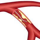
Fractional
Flow Reserve
(FFR) wire in artery
|
|
Dr. Fearon: Like any relatively
new technology, it takes time for people to adopt it and especially,
when data come out, it takes time for that data to get disseminated
and for the technique to start being used.
There's a perception that FFR decreases the number of interventions
and, because physicians are paid for interventions, that
may be a financial dis-incentive. There's also a perception
that it takes extra time to do it and that may discourage
people to use it. But, if we can just keep generating data
showing that it's beneficial and then trying to educate our
colleagues, hopefully with time people will start using it
more and more.
|
Q: Does using FFR really add time to the procedure?
Dr. Fearon: If you have a single vessel and you're deciding whether
to do FFR and stent vs. just stenting, likely it would be quicker
to just stent. But most cases we're dealing with are multivessel
disease and in that setting, we studied that in FAME, and we
found that there was no additional time, that the procedure time
was identical between the FFR-guided group and the angio-guided
group. And likely that's because, although almost every patient
in the FFR-guided arm that got at least one stent, there were
significantly fewer stents placed in that arm and that saved
time, while measuring FFR might add some time, so it was a wash.
I should say that, as people get more experienced using it and
if you have your cath lab set up so that you're prepared to use
it, it's just like doing IVUS. It adds a few minutes, but it's
not like it greatly prolongs the procedure. It's certainly less
than say doing Rotablator.
Q: You mentioned that using FFR results
in doing less interventions, but as Nico Pijls pointed out to
me in his interview, in the case
of multivessel disease, where you might be thinking about sending
the patient to open heart bypass surgery instead of PCI, FFR may
show that only one or two blockages are significant, and so stenting
would be indicated, and you’d actually be adding procedures.
Dr. Fearon: Yes, that's exactly right. That's why I was saying
why it hasn't been adopted more, is that there is a perception
that it decreases the number of interventions. But I would argue,
just as Nico was arguing, that in fact it may add interventions
in some cases, like the one you described.
Q: For those that have taken the plunge and are using FFR, how
has FAME changed their PCI practice?
Dr. Fearon: I think people have really come to realize how limiting
or how misleading the angiogram can be at times. There will be
very modest lesions that in many cases, based on the angiogram,
we would ignore and treat medically and which are actually causing
significant ischemia. And then there are also tighter lesions that
surprisingly are not causing ischemia. I think from FAME and other
studies what we have learned is the importance of identifying ischemia-producing
lesions and treating those and in that way maximizing the benefit
of our stent -- and identifying lesions that aren't producing ischemia
and treating those medically, and therefore minimizing the risk
of putting in stents.
Q: For many patients, who are told that they have a 60% or 70%
blockage -- and they can see a picture of it -- it is sort of counterintuitive
for them to be told that, according to FFR, that is definitely
not an ischemia-producing stenosis. You can see it; you want to
do something to it: it's the oculo-stenotic reflex. But how can
you see something on angiography that clearly looks like a blockage,
but in fact it's not really causing ischemia?
Dr. Fearon: You have to remember that the angiogram is a two-dimensional
image of a three-dimensional structure. You can have a very tight
eccentric lesion that on the angiogram looks very mild. Likewise,
if you're at the right angle, you can have a narrowing that looks
quite severe, yet the lumen is actually quite large. Or even if
the lumen is compromised, the amount of muscle that's supplied
by that vessel is a small amount, so the size of the lumen is adequate
to bring blood to that area. Or the patient's had a prior infarct
in that region, so they don’t need that much blood flow.
There are so many different factors that contribute to whether
or not a patient has ischemia beyond just what the lesion looks
like on the angiogram.
Q: An interesting aspect of FAME was that stenting a lesion that
does not need it, a lesion that's not ischemic, actually seemed
to have worse outcomes, although perhaps not as a cause and effect
thing.
Dr. Fearon: Correct. Certainly some intermediate lesions you can
stent at low risk, but the more stents you put in, the greater
the chance of a complication and what FAME really taught us is
that FFR guidance allows you to more judiciously and accurately
place stents and in that manner maximize the benefit and minimize
the risks of stenting.
Q: At Stanford, do you use
FFR on all of your mutivessel cases at this point?
Dr. Fearon:
Yes. There are certainly instances where you don't need FFR,
like you mentioned, in single vessel disease. If you have a
patient who has typical symptoms and has an abnormal stress
test and you find single vessel disease -- you have all the
information you need there and you can just go ahead and treat
with stenting. But that's certainly that's the minority. In
most cases theres multivessel disease, where you have one
high grade lesion and that is clearly the culprit and you go
ahead and shoot that, but then there's another vessel or two
that has maybe a 50% or 70% narrowing and using the pressure
wire there can be quite helpful. |
|

Stanford
Medical Center |
Q: FAME II is just getting started. What are we going to find
out from this trial that's new?
Dr. Fearon: What FAME II is really going to address again is the
importance of ischemia and its impact on adverse outcomes. COURAGE
compared Optimal Medical Therapy (OMT) for patients with stable
coronary disease to Optimal Medical Therapy PLUS Percutaneous Intervention
(PCI).
What FAME II is going to do is build on that. In COURAGE, the percutaneous
intervention was not guided by FFR, it was an angio-guided study,
so presumably there were instances where lesions that were not
causing ischemia received stents, and perhaps vice-versa, lesions
that were causing ischemia didn't get stented. What we're hoping
in FAME II is that, by using FFR-guidance, we'll be able to identify
truly ischemia-producing lesions and then take that group and compare
medical therapy to stenting. The goal is to look at stable patients
with coronary disease who have real myocardial ischemia and compare
the role of stenting in that setting vs. Optimal Medical Therapy.
Q: So some patients who have ischemic lesions will be treated
by Optimal Medical Therapy only?
Dr. Fearon: Correct. The way the trial is set up in fact is that
in order to get into the study and be randomized, FFR is first
measured. And only if you have an ischemia-producing lesion can
you be included in the study. We're taking an enriched population,
just those with ischemia, and we're then going to compare medical
therapy plus or minus PCI in that setting to try to really get
to the bottom of the benefit of reversing ischemia and the benefit
of PCI.
Q: Fascinating. When can we expect the results?
Dr. Fearon: The follow-up is going to be two years and we started
enrollment just now. We probably won't complete enrollment for
another year or year-and-a-half, so we're looking at three years
off from now, something like that.
Q: In the very beginnings of balloon angioplasty, intracoronary
pressures were extremely important. Gruentzig used to do pressure
gradients on every single patient and really didn't stop the procedure
until the pressure gradient was pretty much eliminated from distal
and proximal. Is FFR harking back to that concept?
Dr. Fearon: Yes. I think the concept of FFR certainly builds on
the early seminal work by Gruentzig and others. One of the big
breakthroughs that Nico Pijls and Bernard De Bruyne made was in
identifying the importance of making these measurements during
maximal vasodilation or maximal hyperemia. The resting measurements
of pressure tend not to be as useful, just because changes in heart
rate and blood pressure and things like that have an impact on
the resting flow, as well as the fact that the heart and the body
are so good at compensating at rest. But during maximal vasodilation,
that's really when the measurements become so useful. The other
key breakthrough was the miniaturization of the pressure transducer
so that these measurements could be made without a large balloon
catheter which could cause obstruction in the vessel. So those
two things were really the key additional things that built on
what was already observed by the early pioneers.
Q: The vasodilation is induced by pharmacological means during
the procedure?
Dr. Fearon: Yes. The most common way is by giving intravenous adenosine.
Q: Do you foresee a big growth in FFR over the next year or two?
Or do you think people are doing a wait-and-see to see what comes
out of the newer studies?
Dr. Fearon: I do think that there's going to continue to be growth.
Again, it takes time for data to get disseminated and become adopted
and for education and for people to learn the technique, although
it's not a very complex technique. So I'm guessing that as that
happens there'll be more use. And then there's ongoing studies
besides FAME II, there are smaller studies comparing IVUS guidance
to FFR guidance. So, as more and more data come out, that will
also generate more interest. And then finally, there are all these
advances being made as far as the wires that are used, and the
technique that make it simpler and easier, and that should help
people adopt it more. Who knows, if maybe there will some changes
in reimbursement too which could always improve its use.
Q: Because of the controversy about
the problems in Maryland and Texas, with doctors now who have
been
accused of over-stenting
and stenting lesions that didn't need to be stented, do you think
that something like FFR could help give some evidence which would
then help justify the procedure. You could say, "Look. This
was necessary. Here's the number."
Dr. Fearon: Right. I think that certainly we're moving in that
direction where whether it's FFR or non-invasive testing or other
methods of assessing for ischemia, but something beyond just the
angiogram needs to be present to justify intervention.
Q: Are you using Fractional Flow
Reserve in other areas that we haven’t discussed?
Dr. Fearon: I think the other area that doesn't get as much attention
is the small vessels, the microvasculature. We focus on the epicardial
vessels, the large vessels, because those are the ones that you
can stent. But there are more and more data emerging showing that,
if we can accurately assess the status of the coronary microvasculature,
that we can have a better idea of the patient's prognosis and also
diagnose the ideology of chest pain more accurately.
The reason I mention this is that with the pressure wire we can
also estimate flow: the pressure sensor can act as a thermistor
and we can measure the transit time of room temperature saline
and get the estimate of flow. And we can use that estimate of flow
in our pressure measurement and then calculate the microvascular
resistance. We've been working on something called the Index of
Microcirculatory Resistance (IMR) -- and we've studied, using this
index, patients who have had acute myocardial infarction, as well
as in patients with stable chest pain syndromes, or patients before
and after percutaneous intervention, and have found that the resistance
in the microvesssels can predict which patients are going to do
well and which patients are going to have big heart attacks and
small heart attacks. As we get more understanding, one could envision,
for example, deciding whether or not to deliver stem cell therapy
based on the status of the microvascular resistance in someone
with an acute MI -- or monitoring the effects of stem cell therapy.
So I think that's another kind of growth area for coronary physiology
and our understanding of the coronary circulation.
Q: Would this be something that's more prevalent in women than
men?
Dr. Fearon: That's exactly right. At Stanford, one of my colleagues,
Jennifer Tremmel, is working on measuring not only FFR but IMR
in women who have normal appearing coronary arteries based on the
angiogram. And we're finding very interesting things. Some patients
have mild diffuse disease that doesn't show up on the angiogram,
yet the FFR is very abnormal. Others have a totally normal FFR
but have a very high IMR, suggesting that they have microvascular
dysfunction or so-called syndrome X. And others have normal FFR
and normal IMR and then you can reassure them that their coronary
circulation is doing fine and likely the chest pain is coming from
another source. So it can be quite helpful and a very thorough
way to evaluate our patients.
Q: How exactly is the IMR measured?
Dr. Fearon: Basically you use the same pressure wire and you again
administer intravenous adenosine and you measure the distal coronary
pressure during maximal flow, maximal vasodilation -- and at
the same time you inject small aliquots (like 3cc) of room temperature
saline. And a kind of thermodilation curve is generated by the
thermistor on the pressure wire and based on that the analyzer
can automatically calculate the transit time. You take the pressure
and divide it by flow and you get a resistance. And that number
is a reflection of the status of the microvasculature.
We found that an IMR of less than 20 is normal and people coming
in with acute MI, usually it's in the 30-40 range -- and the higher
it is, the larger the MI will be. It correlates with the CPK. It
also correlates with the degree of LV dysfunction and also with
the recovery of LV function over time. So that people who have
a high IMR, when you look three months later, their LVs don't improve
as much as those who have low IMR. Hopefully it will become a powerful
tool.
This interview was conducted in September
2010 by Burt Cohen of Angioplasty.Org.
|