|
CT Scans of the Heart Can Be Done with Low Radiation Dose
|
 |
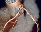
Coronary
artery, as imaged
by a 64-slice CT scanner |
|
February
4, 2009 (updated) -- Physicians
are able to perform high-quality CT angiograms of the heart with
minimal radiation exposure, according to a study published
in today's Journal
of the American Medical Association (JAMA). Using dose-reduction
strategies, some centers included in this study, dubbed PROTECTION
I, were able to perform a 64-slice CT angiogram with a measured
radiation exposure of 2.1 mSv (millisieverts), equivalent
to the level of normal annual background radiation encountered
by a resident of New York
City -- and they were able to do this without degradation of
the image. |
However, some of the 50 international centers
in this study performed similar CT angiograms at ten times that level
of
radiation,
21 mSv,
prompting the authors
to conclude:
"Median doses of CCTA (Cardiac Computed Tomography
Angiography) differ significantly between study sites and
CT
systems. Effective strategies to reduce radiation dose are
available but some strategies are not frequently used. The comparable
diagnostic
image quality may support an increased use of dose-saving strategies
in adequately selected patients."
Interestingly enough, the overall median value
of radiation exposure in this study was 12 mSv which, accordingly
to an editorial in the same issue by Andrew J. Einstein, MD, PhD,
was "somewhat less than the value reported in several
earlier studies using 64-slice scanners". 12 mSv is, in fact, at the lower
end of radiation exposure for the widely-used nuclear stress test --
which can go to 22 mSv for a thallium 201 test.
PROTECTION I was a cross-sectional, international,
multicenter, observational study of 50
sites,
including 21 university hospitals
and 29 community hospitals. The team, headed by Dr. Jörg Hausleiter
of Munich, looked at the estimated radiation dose in 1,965 patients
undergoing CCTA between February and
December 2007. PROTECTION I is one of the first studies to look at and measure
actual radiation dose from CT heart scans. The important finding
is that there is
very wide variation among centers; the important message is that
there
are
strategies
for reduction
of radiation
levels
for
cardiac
CT angiograms, and that centers performing these tests need to be
aware of and trained in these techniques.
According to Dr. Tony DeFrance, Medical Director
of CVCTA Education in San Francisco:
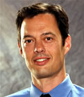
Tony DeFrance,
MD
Medical Director
CVCTA Education
San Francisco, CA
|
|
"We need to get the
word out to the centers and I think education, ongoing dialogue,
even site visits for people are important to optimize their
radiation
reduction strategies. Step one is measuring their doses. What
weve done, and what's really changed our practice, is measuring
the dose on every patient and recording it. That's the first
step to awareness: knowing how much dose you're
delivering. Then getting educated about the different dose
reduction
strategies, from dose modulation to prospective-gated imaging.
About a year-and-a-half ago, we were getting an
average of 16 mSv. Since we've instituted
our dose reduction strategy our average dose now is 6.8. It's
just a matter of doing prospective imaging as much as possible,
using dose modulation
and really optimizing the scanner technology and parameters to
do it. I think a lot of centers aren't using them -- they're
just using retrospective
on everyone. So the key
is teaching these people that you can halve the dose or even
decrease
it by 80%. |
Dr. DeFrance and CVCTA Education use the acronym
LOWDOSE to summarize what CT centers need to think about:
Length of scan minimized
Optimized X-ray parameters and gating technique to the individual
Working with vendors to optimize the system
Dose Modulation
Operator Training (CT Technologist and physician)
Scrutinize the effective dose on each case
Evaluate dose at regular intervals and implement changes to minimize
it
Implications for Patients
The PROTECTION I study also has significance for patients who
are scheduled for a CT angiogram. Dr. Michael Poon, past president
of
the Society
of Cardiovascular Computed Tomography (SCCT) and Director
of the Center of Advanced Cardiac Imaging at Stony
Brook University Medical Center, told Angioplasty.Org:
"The patient has to request
this. Patients may read about this, may see the headlines,
and then
they need to go back and ask the imaging center, 'What method
are you using to lower the dose?' And if they don't know what
you're talking about, I would say, 'See you later!'
"I'm very
serious. I'm doing that at Stony Brook. I've told the hospital
that when the patients come, I say, 'Look, I am doing this
with the lowest
dose possible. If you can find a lower dose then go there.'
But they can't, because I know the method, I know what it takes,
I
know what to do to get the patient into the range so that you
can take advantage of this latest technology to get the lowest
dose." |
|

Michael
Poon, MD, FACC
Stony Brook University
Medical Center |
One of the other caveats in interpreting the results
of the PROTECTION I study is that it used data from the year 2007,
which does not take into account the extensive dose-reduction protocols
and equipment advances from all the major manufacturers of CT scanning
equipment that occurred in the past year,
from GE's prospective
gating
to Toshiba's
320-slice
scanner and similar
programs for Philips and Siemens, so the radiation exposure is most
likely lower than what is reported. Dr. Poon stated,
"The article
is not representative of the latest technology that we are using."
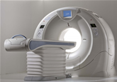
Aquilion ONE
320 detector row
CT scanner |
|
Dr. DeFrance notes that
much incorrect information is being distributed. For example,
many people might think that Toshiba's 320-slice scanner
produces excessive radiation, compared to a 64-slice machine.
But, in
fact, the opposite is true because it is able to scan an entire
heart in one gantry rotation of 300 milliseconds. Dr. DeFrance
currently does CT angiograms, achieving radiation
doses of only 1 or 2 mSv.
Similarly GE's new high-definition CT system
is able to image a very high quality at low exposures -- neither
of these technologies were in wide use during the time frame
of the PROTECTION I study. |
The conclusion and rationale for this study
is that, with training and education, CTA radiation levels can be
significantly
reduced. Yet many news reports of the study only emphasize the highest
radiation levels measured. Dr. DeFrance opines:
"There's so much misinformation
out there. And CTA is such a good modality. I think we're
losing
the media war. There are a lot of people with vested interests
that don't want this to succeed. And new technology adoption
is center-focus
for CMS, for the new Administration and, in how to bring a new
technology with comparative effectiveness, CTA is the frontline
battleground
of this. What always bothers me is that the media throw these
numbers around, but what they don’t
realize is the risk-benefit ratios and the risk of coronary disease
vs. the
theoretical risk of cancer, they don’t
even compare it, and the articles don’t even address that."
An important point, emphasized by all cardiac CT
practitioners, is that CTA is not a
screening tool. It is
optimally
used for
patients
who are experiencing symptoms of angina, but in whom other tests
have proved inconclusive. Even at low dose, CTA is able to accurately
rule out coronary artery disease (CAD). The alternative is to send
a patient for an invasive cardiac catheterization, which is significantly
more
expensive, which exposes the patient to the risks of an invasive
procedure and which 37% of the time results in a negative finding
for CAD, one that could have been detected much more simply by using
CTA.
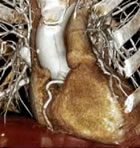
See
a "CT Tour of the Heart"
by Dr. Harvey Hecht |
|
About the Imaging
and Diagnosis Center on Angioplasty.Org
Founded in 1997, Angioplasty.Org is the Internet's most popular site
devoted to interventional procedures. Imaging of the coronary anatomy
is fundamental to the diagnosis and treatment of coronary artery disease.
The Imaging and Diagnosis Center was created to communicate
the most current developments in this field to both patients
and healthcare professionals, featuring interviews with innovators
in the field, news, a discussion forum and a "Patient
Guide to Heart Tests".
For more information, visit Angioplasty.Org's Imaging & Diagnosis
Center.
|
Reported by Burt Cohen, February 4, 2009
|