
|
Multislice
or Multidetector 64
Slice CT Angiogram (2007 and earlier)
|
|
Have you had a multislice CT angiogram
(Cardiac CT) to check for blockages? Did you continue on to a
catheterization or was that not necessary? Post your comments
or questions about Cardiac CT to this topic. Other postings can be found here: Current Add Your Comment See More Topics |


|
|

| More from the Forum: |
| • Browse through existing topics on the Patient Forum |
| • Create a New Topic on the Patient Forum |
| Click here for more information about the following ads |
Find out more about this topic in our feature, Multislice CT Angiograms.
Archived Postings from 2007 and Earlier (109):
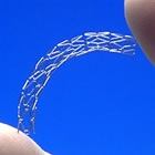
• Jane -- visualizing stents with CT angiography
is tricky. The newer more sensitive 64 slice systems are pretty good, but
there are a lot of variables. What kind of problem with the stent did the
CTA show? As you can read in our special section on Intravascular
Ultrasound,
even invasive angiography (catheterization) can miss whether or not a stent
has been perfectly implanted. We're assuming, from your post, that you
are not covered by insurance, but a potential problem with a stent should
be dealth with. Perhaps you could talk to the doctors involved (the interventional
cardiologist who did
the
stent
and the
CT reader)
and
ask
them if they would agree to a quick consult at a reduced fee?
Angioplasty.Org Staff, Angioplasty.Org, January 5, 2008
• I am a 53 year female. At 51 I had a cardiac catheterization after a myocardial
infarction. My LAD had eccentric stenosis of 60% in mid segment after diagonal
branch. The LAD was totally occluded distally with thrombus. AngioJet thrombectomy
on thrombus and distal LAD was successful, however I still continued to have
distal occlusion near the apex. A 2.5 x 23mm Cypher stent that wrapped around
the apex was deployed in order to establish a better distal flow. Recently my
regular doctor ordered a CTA. He told me that the doctor that read the CTA found
a problem with the stent. He made an appointment with me to see the doctor that
put the stent in during the catheterization. Because of financial problems I
am unable to afford the visit to see the doctor that put the stent in. Should
I be concerned about trying to get checked for this concern my regular doctor
told
me about after the results of the CTA?
Jane, Ohio, USA, December 29, 2007
• I have had several coronary stent procedures
and am interested in assessing them for restenosis as I have had this
occur 2 yrs ago. I am interested in obtaining a ct angiogram. However I
have
some renal insufficiency (serum creatinine 1.8 and bun 29). If I decide
to obtain the ct study what would be your pre-study recommendations to
prevent contrast nephropathy i.e. acetylcysteine, etc., or should I forego
the ct study?
Harold, Colorado Springs, Colorado, USA, December 19, 2007
• Craig -- your cardiologist is correct -- a high Calcium Score by itself doesn't indicate coronary artery disease (CAD) -- and most 16 slice CT scans don't have a high enough sensitivity to see soft or "vulnerable" plaque. What mutlislice, or 64-slice CT angiography is excellent for is to rule out CAD, definitely. There is a consensus statement that was authored by the major heart societies (AHA, ACC, SCAI, SCCT). It took some time to put the guidelines together because unnecessary testing is an area of concern for all. Additional radiation from the 64-slice CT is also a concern, but it's a bit less than what you'd be getting from a nuclear stress test and, with newer equipment and methods, it's far less. If you read some of the interviews with imaging experts in our Imaging and Diagnosis Center, you'll see one clear difference between stress tests and a CT angiogram. The stress test is a functional test -- it shows if the flow of oxygenated blood to the heart is less than optimal. It shows a result, but not the cause. A CT angiogram is a direct view -- it will show if there is are no blockages with 99% accuracy. We can't advise you one way or the other. This is a conversation you should have with your cardiologist and you should discuss the cost/benefit -- not forgetting that putting your mind at ease would be a benefit. One caveat -- CT angiograms are a bit less accurate when in the presence of a lot of calcium, which acts as a shield.
And Krystal -- an infarct would mean that at some point
your husband had a heart attack and some of his heart muscle is damaged
-- your husband is very young for such a diagnosis.
EKGs
are
the
first line of testing and are by no means a test on which a firm diagnosis
can be made. But an abnormal EKG should
lead to additional testing. Discuss this with your doctor for sure.
Angioplasty.Org Staff, Angioplasty.Org, November 12, 2008
• My Husband is 24 and has history of heart problems.
He recently had a EKG done and it is reading abnormal. He has several of
them and they are all abnormal. It reads Interior Infarct. I would like
to know what that means and
what I should do? Thanks.
Krystal, North Carolina, USA, November 9, 2007
• 52yr/M/asymptomatic/ My father had a triple bypass
in 1973 at 48 years age and lived another 30 years. My wife is also
a heart nurse. For that reason I had all the stress tests done three years
ago.
I am an amateur climber and do a great deal of exercise, mainly Stair
Master and hiking uphill with heavy loads, 40 to 65lbs. I am very fit.
My stress
test indicated no problems. I then asked the cardiologist about a calcium
scan, which at the time was an 8 or 16 slice. He laughed and said not
to get paranoid because I was fine. He said I simply could not do what
I did
on my nuclear stress test if I had any problems. I did the calcium
scan
anyway and scored about 260 which put me in the 90th percentile. My
blood work was good and my cholesterol was normal. Even so the cardiologist
put me on one baby aspirin per day and zocor. I have been on this regimen
for
three years. Now the Heart Hospital has the 64CT scanner and I am wondering
if someone who is asymptomatic but had a high calcium score should
get
the 64CT angio scan? My cardiologist told me hard calcium wasn't what
I should worry about. He said it was the soft plague that gets you in
trouble and the 16 slice didn't pick up soft plaque. What is the current
day
consensus
about an asymptomatic person who is very fit but has a high calcium
score, undergoing the more comprehensive
64CT angio tests?
Craig Blankenship, Edmond, Oklahoma, USA, November 8, 2007
• Joan -- we are not in a position to either offer
medical advice or to second-guess the advice of a qualified MD. If
you are concerned about whether you should get a CT, you can browse through
the "Appropriateness
Guidelines for CT", authored by the American College of
Cardiology as a guide for when a Cardiac CT is indicated. You will
note a complex scoring system which measures the various trade-offs,
one of
which is the radiation dose
from a
typical
Cardiac
CT - roughly
equivalent
to what you would get from a standard invasive angiogram. One question
to ask your physician is how will the results of this test affect any
course of treatment?
Angioplasty.Org Staff, Angioplasty.Org, October 7, 2007
• My doctor wants to do this as I have a family
history of early onset CAD. I am 54 and I have no symptoms or any other
risk factors. My CT heart scan 3 years ago was excellent--no calcium,
but he says I could still have soft plaque. An echo stress test was good.
VAP
test was pretty good. He'd like me to take plant sterols and fish oil
to get my LDL from 82 to the 70's, and to change the small dense LDL pattern.
I have tested negative for inflammation. What concerns me is the radiation,
the possible reaction or ramifications from the iodine dye and serious
(though rare) reactions to beta blockers. Thank you.
Joan D, Denver, Colorado, USA, October 3, 2007
• Reimbursement for CTA is a big issue. It has changed
and will continue to change as more studies are published showing its accuracy.
The major cardiology associations have updated their guidelines
for use of CTA and the SCCT,
which specifically deals with CT, is active in advocating for increased
reimbursement. You might want to contact them directly. We're curious --
what was the result of the CTA??
Angioplasty.Org Staff, Angioplasty.Org, October 3, 2007
• I had a CTA three months after having a cardiac
catheterization and a stent placed in my anterior descending artery.
The need for the CTA was persistent angina following the placement of the
stent.
Blue Cross Blue Shield of Michigan is refusing to pay for the $1800.
procedure (They categorize as investigational). BSBS would have paid for
another
cardiac catheterization that would have cost several thousand dollars
more! Any suggestions on how to get this paid, different
coding etc.?
Gary C., Michigan, USA, October 3,
2007
• Vy -- you are correct. These is no femoral
or radial arterial puncture done in a CT Angiogram -- only an IV. But
definitely let your CT doctor know about your Coumadin prior to the appointment
in
case they have a specific protocol to follow.
Angioplasty.Org Staff, Angioplasty.Org, October 2, 2007
• I am scheduled for a CT Angiogram due to a chronic
pulmonary embolism. I am on Coumadin for this condition. My hemotologist
thinks that this procedure would make it necessary to stop Coumadin for
a few days before the procedure
because "Any kind of angiogram involves an incision" IS this true? Isn't this
procedure done with a conventional vein IV?
Vy, California, USA, October 1, 2007
• ST -- beautiful mountains! Wish we were there.
A CT Angiogram (64 slice) has been shown to be highly negative predictive
(99+%).
In other
words, if the CT shows no disease, you can pretty much trust it. It's
a bit less accurate in showing positives, because there can be false positives
due to a number of things -- although it's still pretty accurate, and
getting
moreso all the time.
Angioplasty.Org Staff, Angioplasty.Org, September 17, 2007
• I am a 55 yo postmenopausal female who hikes vigorously in the back country
of Glacier National Park. My stress test yesterday yielded ST depression. I will
soon be undertaking a CTA. Is this the logical next step or what other possibilities
exist? How definitive is a CTA?
ST, Montana, USA, September 14, 2007
• What is the upper permissible limit of radiation
a patient can receive in his/her lifetime? How many times in a year a
patient can be exposed to the radiation?
Amruta, Siemens, Bangalore, INDIA, September 11, 2007
• Betty -- 64-slice CT actually uses a similar dye
to standard angiography -- Magnetic resonance does not. Although if you
might have renal insufficiency, make sure that gadolinium-based dye is
not used in your MR. There are non-iodine dyes and also precautionary protocols
to pre-treat people who are allergic to iodine-based contrast. We can't
recommend a hospital -- perhaps a query to the Society
for Cardiovascular Computed Tomography (SCCT) would help you find a qualified imaging center.
Angioplasty.Org Staff, Angioplasty.Org, August 14, 2007
• I have hypertension that has become difficult
to control, my cardiologist wants me to have renal and cardiac catheterization.
I am allergic to iodine dye and was wondering if CT or MRI would be better
for me to have. I understand the 64-slice CT requires no dye. I live
in Arkansas and would also like to know the nearest hospital where I might
have this done. I can travel somewhere else if it is safer for me to
have.
Thank you, Betty Ellis
Betty Ellis, Newport, Arkansas , USA, August 12, 2007
• Hi, I'm the proud new owner of a stent (1 week).
I'm 50 and had NO symptoms. I did a coronary calcium scan (had to pay $600
myself, as I am in the U.S. where insurance won't cover it) and discovered
significant calcium deposits in my LAD artery. I pushed my cardiologists
for more testing. My Manhattan cardiologist just gave me the conventional
stress test, which I passed with flying colors. I pressed for a nuclear
stress test which he balked at. Mind you I had: 1. Family history of heart
attacks, 2. Very low HDL score, 3. High tryglycerides, 4. and a coronary
calcium score very high for someone my age. NOT good enough for an insurance
company to pay for more testing! Finally switched to a new cardiologist
and just happened to develop chest pains that migrated to my arm, so mild
that I normally would have thought mild heart burn. But with the calcium
sore I decided to make a BIG deal about it. I got an angiogram. Sure enough-
80% blockage! Medicated stent put in place and I feel the improved circulation
already! What's the lesson? You have to be assertive and your own health
researcher. In the US the insurance companies would rather I have sudden
cardiac death. That way I cost them nothing. The doctors can be too intimidated
by the insurance companies to be your advocate. At least this was the case
with my Manhattan cardiologist. The cardio that prescribed the angiogram
seem really humbled when he saw the results.
John, Nyack, New York, August 4, 2007
• Darlene -- the contrast dye used for a 64 slice
CT angiogram carries the same cautions as the dye used in a standard
invasive angiogram. However, there are dyes that are less toxic to the
kidneys --
and in some
cases, the dye can be diluted more than usual, but you need to have a very
good
imaging system to get the best resolution, whether you go CT or invasive.
It makes no sense to subject a patient to any test if the test is not going
to provide
the
proper
information.
Your situation is not uncommon, a significant percentage of patients with
coronary
blockages
also have
blockages in the renal artery, causing kidney dysfunction -- and vice-versa.
Cardiologists who do these procedures have a number of protocols in place
to protect the kidney (keeping the patient super-hydrated, etc.). Your
nephrologist should discuss these issues with your husband's interventional
cardiologist
to
make sure
every protocol is followed. The CT angiogram is definitely less-invasive
and may be the appropriate test if your husband is in that grey "indeterminant" area.
However, this is a decision that needs to be made by you with both of
your doctors. If there's a good chance your husband is going to need
an invasive angiogram anyway (or angioplasty/stent) then it would be best
to go right to that test and lessen both the contrast dye and radiation
exposure.
Angioplasty.Org Staff, Angioplasty.Org, July 22, 2007
• My husband , age 72, is diabetic with some kidney
impairment. His cardiologist wants to perform an angiogram because of his
stress test results but is leaving
the decision up to my husband and his nephrologist. Would a 64 slice CT involve
less contrast and lessen the amount of damage caused by the contrast dye?
Darlene K., Arizona, USA, July 18, 2007
• Annie -- it would be good if the cardiologist
who did the invasive angiogram could contact the radiologist who did the
CT and perhaps it could be a learning experience for both. We're awaiting
the results of the CorE 64 trial in the fall, which pits 64 slice CT
angiograms against the standard invasive angiogram -- but studies done
with 16 slice units show a very high negative predictability: if the CT
shows no disease, there is none. 16 slice fell down a bit on positive predictability,
but it was still pretty accurate -- mainly not doing as accurate a job
with intermediate blockages. Not sure what to say about a severe blockage
turning out to be none at all. We would urge all posters to read through
the various articles on our Imaging and Diagnosis Center, especially our
current interview with Dr. Stephan Achenbach, who addresses some of these
issues.
Angioplasty.Org Staff, Angioplasty.Org, July 18, 2007
• A month ago I had the 64 slice CT Scan I paid
for myself.It was a preventative due to family history. I had all the
stress tests and a battery of other things for a few bouts of chest pain.
This
week I had an angiogram. Reason, the CT scan radiologist said I have
a severe blockage. When the Doc went in he said NO blockage. Radiologist
must have read it wrong! Why I am told my heart is the less than 5% that
has a right artery that enters different spot on my heart. Why could
they
not see this??? I am suspicious now. Also I am told the normal heart
has pressures around 5cm and mine is 30cm. I have used nitro for severe
chest
pains now on Avalide 150/12.5 Metoprolol - Toprol XL 25 mg 81 mg aspirin
So what is
interior heart pressure and how do you fix that?
Annie, Ohio, USA, July 15, 2007
• Rick -- do you mean "should I be thinking about
an angiogram"? An angioplasty is a treatment for a blocked artery, and
so far tests have shown you have none. Have you had a Calcium scoring exam?
This involves much less radiation than an all-out 64 slice CT angiogram,
and might help relieve your concern. Were your brothers' heart attacks
caused by a blocked artery? Or by a rhythm dysfunction? Runners and other
atheletes sometimes develop a slightly thickened chamber wall in the heart
which masks cardiac rhythm problems. Angiograms test for arterial blockages.
Angioplasty.Org Staff, Angioplasty.Org, July 15, 2007
• My two younger brothers, ages 49 and 51 respectively
have had heart attacks, the younger one just a few days ago. After the
older one had his which he survived
with 5 stents to deal with his 95+ blockage, I had a nuclear stress as well
as a sonogram, both of which were negative conducted by my cardiologist.
Now the
younger one, a former world record holder in distance events just suffered
his heart attack a few days ago and also survived. He would appear to be
in excellent
shape with daily workouts and not a likely candidate compared to me, his oldest
and much heavier brother, age 53 but also a former runner. I have had no chest
pain but a two instances of almost passing out that weren't fuly explained
along with several of lightheadeness and just not feeling right that have
lasted from
several hours to a day or two during the last year. Should I seriously be thinking
about a angioplasty?
Rick, Tehachapi, California, USA, July 3, 2007
• Glad to hear things went well. Sometimes (not
always) our own sense of our body is one of the best tests -- your sense
that something was amiss (even thought the Thallium stress test had a
good result) led you, along with your cardiologist's recommendation, to
the
correct treatment. It's odd that there was no indication of a perfusion
deficit (reduced flow) during the stress test, especially since you had
a 90% blockage -- but it is possible that the blockage increased in the
time since the test. Had you been having chest pain for a long time,
or just the past 6-12 months? Genetics are a definitely strong factor.
We
know a story about twin brothers -- one died suddenly from a coronary
blockage, so the twin decided to get a cath. Turned out he had a lesion
(blockage)
in exactly the same spot in the same artery as his less fortunate twin.
As for how to monitor your future health, by all means discuss this with
your cardiologist. CT scans are more readily available than cardiac MRI,
but they do involve radiation exposure. And both imaging modalities are
rapidly changing, getting more accurate and advanced with time. As for
radiation from a multislice CT -- a 64 slice scan, especially if done
in a gated fashion which can reduce radiation exposure, is not different
in
the amount of radiation than a Thallium stress test -- and may even be
less.
Angioplasty.Org Staff, Angioplasty.Org, June 1, 2007
• I just thought I'd follow up on your (the Forum
Editor's) response to my original post of May 24. I appreciated your discussion
of testing alternatives. I decided to undergo the angiogram which I did
on May 25 and which resulted in the discovery of a 90% plus blockage and
placement of a 3.5 x 28mm SES stent in my LAD. I am feeling well and expect
to fully recover. I am writing to flesh out some details in the hope others
may benefit. My decision to undergo catheterization was affected by an
episode on Thursday (5/17) including chest pain, pain in lower jaw and
cheeks and lightheadedness. The episode lasted about 40 minutes. I had
had somewhat similar episodes (maybe two or three a year)since 2001 but
they never lasted more than 7 or 8 minutes. In addition, I had been to
the ER after one such event in 2002 and again in 2006.No heart problems
were discovered on either occasion. Furthermore, after the 9/2006 event
I had a thallium stress test which I passed with flying colors. So, I didn't
think any of the previous episodes including the one on 5/17/07 was heart
related. Being in excellent condition, I continued to think--- heartburn.
In fact, that same afternoon following the event I did my usual 45 minutes
on the stairmaster with no discomfort. However the 5/17 symptoms were more
pronounced and lasted far longer than ever before. This scared me and in
conjunction with my bad family history prompted a decision to let my cardiologist "cath
me up" to use his vernacular. It turned out to be a wise choice. But now,
for obvious reasons, I am reluctant to rely on stress testing in the future.
Do you think that I should be requesting a cardiac MRI or multislice CT
scan every year or two to monitor my heart?
Richard S., North Carolina, USA, May 30, 2007
• Richard -- how to decide? That's the 64 dollar
(or 64 slice) question. There are two types of diagnostic tests: visual
tests that show blockages (angiogram, whether invasive or CT) and functional
tests that measure blood flow quantitatively (stress tests, FFR, perfusion
imaging). Current research is ongoing and soon a CT scan may be able to
accurately do both types of tests. Your doctor is correct about the "gold
standard", but that opinion is in flux right now. Invasive angiography
has long been considered the "gold standard", but in a recent
discussion we had with Dr. Harvey Hecht of Lenox
Hill Heart and Vascular Institute of New York, he opined that multislice
CT was really the new gold standard and actually yields more information
if done correctly. (Dr. Hecht is not only an imaging expert, but began
working in this field with the pioneers of angioplasty.) Most cardiologists
we've spoken to feel that the clear role of multislice CT in diagnosis
is for "intermediate" cases, where the results of a stress test
are inconclusive and where the likelihood of coronary artery disease is
not high or low. This is also currently the recommended guideline. If your
likelihood is high, then you may need to get an invasive angiogram anyway
-- and by doing both tests, you'll be doubling your radiation dose. Another
reason for going directly to an invasive angiogram is that, if a significant
blockage is found, it can be treated via stenting in the same session --
sometimes called "ad hoc angioplasty". However, some cardiologists
feel that this linking of diagnosis and treatment has led to overuse of
angioplasty and stenting. On the other hand, if your risk factor is intermediate,
why get an invasive angiogram (which does have risks, although small in
number) when a 10 minute multislice CT scan will yield the answer. One
always has to ask, how will this test change my treatment? So, the best
course of action is to consult with a cardiologist who can look over your
history and discuss this with you, and help you make the best decision
for your particular case.
Angioplasty.Org Staff, Angioplasty.Org, May 24, 2007
• Hi. I am a healthy 57 yr old who exercises regularly.
However several males on both sides of the family died in their early 50's
from heart disease. In addition, my brother ,who is a year younger, had
a heart attack and Angioplasty at age 37. After I experienced chest pain
my doctor recommended a catheterization, which he described as the "gold
standard" for analyzing blood flow to the heart. I don't want an invasive
procedure if the cardiac MRI or Multislice CT will give as good (or almost
as good ) a picture of my problem. My question is--how do I choose?
Richard S., North Carolina, USA, May 24, 2007
• I had a CT scan last year and have since received
3 angiograms and 3 DES stents. My doctor wants another angiogram but I
d like to consider a 64 bit scan. I also have two younger brothers both
experiencing chest symptoms similar to mine. And like me, they have both
been given a clean bill of health on all three types of stress tests. I'm
recommending a scan of sorts as my CT scan prompted my doctor to get me
in for my first angiogram where they found a 99% blockage. I would like
some feedback on your experiences, results and cost.
ASF, California, USA, March 12, 2007
• Karen -- there's no question that the 64 slice
CT angiogram is less invasive. An IV of contrast dye and 15 minutes later
it's done -- no puncture of the femoral artery, no catheters, arterial
complications, etc. It's also less expensive, but obviously not if you
have to pay for it yourself. Reimbursement for Cardiac CT is a big issue
right now -- especially as more and more studies are published showing
its diagnostic accuracy. In your case, the big question is for your interventional
cardiologist or surgeon -- and that would be whether they think they can
get the information they need from the CT. As far a blockages go, the 64
slice CT has been shown to be highly accurate in excluding coronary artery
disease. In other words, if the CT shows no blockage, then there probably
isn't any. But there may be other things that your doctors are looking
for, so this is a conversation you should have specifically with the doctor
who would be doing any treatment. Normally an invasive angiogram is recommended
if it is thought that treatment is likely and would be done in the same
session. The radiation dose for a multislice CT is similar to that done
in an invasive angiogram, although this can vary widely, depending on how
the CT is done.
Angioplasty.Org Staff, Angioplasty.Org, March 9, 2007
• Hello, I am a 35 year old female in relatively
good health. I had open heart surgery on May 8, 2006 to ligate a RCA to
LV Coronary Artery Fistula which was misdiagnosed when I was a child as
a heart murmur. I developed a cardiac tamponade, which required a second
surgery on May 13, 2006. After being released from the hospital, I developed
an infection in my incision (which was caught early and taken care of with
antibiotics), I then caught pneumonia which was also treated with a course
of antibiotics. My sternal incision has formed a keloid scar, for which
I have received injections of kenalog to minimize the pain & itch(3 injections
to date). I was recently (1/23/07) hospitalized for chest pains and had
several tests. The cardiac cath done prior to CAF ligation revealed no
blockages, but the tests done during the Jan 2007 hospital stay were as
follows: ECG returned an inferior infarct, X-rays revealed an enlarged
heart, nuclear stress test indicated probable issues with front & back
of heart. The doctors also suspect that my sternum hasn't fully healed
due to the rocking and crunching that happens when they palpate my chest.
One of the cardiologists I saw recommended that I undergo another cardiac
cath. I was hesitant due to the complications I had following the surgery.
I obtained another opinion, and was advised that I could choose to have
a 64 slice CTA scan performed in lieu of another cardiac cath. My quandary
is that while my insurance will cover a cardiac cath, it will not cover
a CTA scan. I would gladly pay for the CTA out of pocket if this will accurately
diagnose what is causing the chest pains; however, if I will have to eventually
undergo a cardiac cath anyway, I would rather skip the CTA even though
I am hesitating to go with another invasive procedure. Any advice would
be appreciated!
Karen A., Hawaii, USA, March 8, 2007
• Does anyone know the codes for insurance purposes
for this procedure -- Multislice CT Angiogram -- (diagnosis code and procedure
code)? I am trying to find out if my provider covers it - Highmark Blue
Cross/Blue Shield. Thanks
John Burkovich, Monroeville, Pennsylvania, USA, March 7, 2007
• Eileen -- we assume you mean Calcium scores --
which are done without contrast and at a low radiation dose on either a
multislice CT or an electron beam CT. They range from 0 to over 400, with
zero signifying virtually no calcium deposits, and anything over 400 being
high. A number of studies show a correlation between calcium scores and
coronary blockages (they are not the same thing -- it is possible to have
one without the other). A significant calcium score, along with an ultrasound
of the carotid artery showing plaque, has been identified in the recent SHAPE
guidelines, as an important indicator of coronary artery disease, one
which would point to further testing, either with a full dose 64 slice
CT that could show arterial blockages, or an invasive coronary angiogram,
done in the cath lab.
Angioplasty.Org Staff, Angioplasty.Org, March 7, 2007
• I recently had a 64 slice Cardiac CT and was told
I got a score of 1. What does that mean?
Eileen, New York, USA, March 6, 2007
• Patrick -- thanks for the clarification. Our Imaging and Diagnosis Center describes the various tests given cardiac patients. The nuclear stress test doesn't yield the kind of visual picture of the blockage that one would get from an angiogram of the artery. It is what's called "a functional test" -- that is, it measures the blood flow to a specific part of the heart. For example, an angiogram might show a 50% blockage, but the important question would be "is that blockage an obstructive blockage? does it actually restrict blood flow and cause ischemia to the heart?". To answer that, one needs a "perfusion study", which is what a nuclear stress test is. It measures how much blood flow is perfused through the artery to the heart muscle. If a nuclear stress test is inconclusive, then the next step is usually an angiogram. There is thought that within the near future, the nuclear stress test may be replaced by the multislice CT angiogram -- in our interview with Dr. Armin Zadeh, he discusses research now being done at Johns Hopkins that would allow the CT to be both an angiogram AND a perfusion study simultaneously: a "two-fer" in effect.
By the way, the fact that the stent in your RCA was large
is good (we assume he meant wide) -- large diameter arteries definitely
have lower restenosis rates. As for the fact that your insurance wouldn't
cover Plavix the whole time, the recent "Joint
Science Advisory", issued by all the major heart organizations,
concludes with calls upon Congress and insurers to prevent just such a
situation. Finally, regarding your other stents that are closed "like
concrete", there are new technologies being developed to open what
are called CTO's -- or chronic total occlusions. There is in fact a major
meeting going on today and tomorrow in New York, called the "Chronic
Total Occlusion Summit", addressing these issues. But these are complex
situations and, as we always advise, your cardiologist is the one who knows
your clinical status best, and also is intimately involved with what is
or is not possible, or even helpful. (Just because he "can" do
something, doesn't mean he "should".) What you "should" do,
however, is whatever you can to reduce your risk factors, which you probably
already do -- keep cholesterol and blood pressure under control, no smoking,
keep as active as possible, etc.
Angioplasty.Org Staff, Angioplasty.Org, February 22, 2007
• Please let me clarify. After my nuclear stress
test this January, I had another angiogram and had a stent placed in the
RCA, which was now 90 per cent blocked. The cardiologist said it was a
large stent. He told me that the first stents had completely blocked "like
concrete" and they couldn't be opened. I have been on Plavix and Aspirin
the whole time, except for brief periods when insurance wouldn't cover
it and when I had surgery- just a few days. This procedure was earlier
this month, on February 2nd. My concern is that if this new stent blocks
up like the old one, I will die. If a nuclear stress test can not measure
how much blockage there is, then how do I monitor the situation? Exactly
what can be seen by a nuclear stress test anyway? Can't they just do an
annual heart cath angiogram?
Patrick, Costa Mesa, California, USA, February 21, 2007
• When you say "the cardiologist said it couldn't
be opened") was this an interventional cardiologist? We're not clear
how it could be determined that you had a 100% blockage via a nuclear stress
test? The stress test shows function -- it can measure ischemia -- but
it doesn't visually show a blockage. One reason to have a cardiac cath
or standard coronary angiogram in cases like yours is that it is possible
to open up a blockage if one is found. By the way, nuclear stress tests
can involve a not-insignificant amount of radiation. CT angiograms also
involve radiation -- that amount is being reduced as new methods and technologies
are introduced. And a CTA can show a blockage -- however, if there is evidence
of blockages and restenosis (reblockage) a cardiac cath may be the best
diagnostic test -- mainly because it can quickly be converted into a treatment
in the same session.
Angioplasty.Org Staff, Angioplasty.Org, February 21, 2007
• I am a 47 year old male. I have several negative
risk factors working against me. My father died at 74 of heart failure
after having at least 3 heart attacks and a few open heart surgeries, starting
when he was 51. I had an MI in January of 2004 and had 2 drug eluting stents
placed in the LAD artery. Then, the RCA was 60 per cent blocked. I pushed
for a nuclear stress test, and had one this January. My stents were 100
per cent blocked (the cardiologist said that it couldn't be opened) and
the RCA was 90 per cent blocked. I was told that if it reached 100 per
cent, I would die. He recommends a nuclear stress test every year. Some
collateral arteries are now providing blood flow to the left side from
the RCA. I have been on Plavix all the time since the first angiogram in
2004. Are there other ways of monitoring any blockage in the new stent
in the RCA, my life literally depends upon it.
Patrick, Costa Mesa, California, USA, February 15, 2007
• Sharel -- chest pain is not always inidcative
of coronary artery disease. And in women, chest pain is a bit more complex.
Of course, your doctor is the best source of advice on any of these questions,
but we would assume that since all of your mother's tests showed negative
for ischemia and that she has been judged at low risk for coronary artery
diease, medical management and life-style changes are the way to go (we
assume she doesn't smoke, etc.). A CT Angiogram varies in cost -- you can
call an imaging center to verify -- but the question is whether there is
a reason to get the test (there is also a radiation dose involved). CTA
is very useful and accurate in ruling out CAD in intermediate cases. But
you should discuss this with your mother's cardiologist. We think he/she
would agree that a standard invasive angiogram is certainly not indicated.
Angioplasty.Org Staff, Angioplasty.Org, February 14, 2007
• My mother (58yrs) went in to the hospital a few
days ago for chest pain, and high BP. SHe had been on medication for high
BP for several years. She was admitted in the hospital. Her EKG was normal
at first but troponin level was high. After a few days, troponin fluctuated
and EKG showed dynamic changes. Because her blood was not flowing well
on collection, she was put on heparin intravenously and diagnosis was Coronary
artery disease. Cholesterol is normal. After angina and burning sensation
in chest subsided, her ECHO and stress test showed negative. Her diagnosis
now was no ischemia or not at major risk. She has been released with aspirin,
increased dose of beta blocker (atenolol) and lipitor. She was told an
angiogram was unnecessary. Based on her tests. Is an angiogram advisable?
What is the out of pocket cost?
Sharel, Irvine, California, USA, February 12, 2007
• David -- because everyone's clinical situation
is different, we can't recommend a specific test or treatment for any patient
-- that's up to your cardiologist. But you can definitely ask him/her why
they recommend a standard angiogram and not a multislice CT -- they may
have a very good reason and you'd be more confident if they could tell
you why, in your particular case. One possible reason would be that there's
a likelihood of an intervention being necessary, which can be done during
a catheterization, but not during a CT angiogram, which is non-invasive
and not done in a cath lab. On the other hand, state-of-the-art 64 slice
CT angiograms have a very high (99+%) negative predictability -- that is,
the CT is very accurate in ruling out coronary narrowing. Does this hospital
have a multislice CT scanner?
Angioplasty.Org Staff, Angioplasty.Org, February 10, 2007
• Had an eluting stent placed in LAD 18 months ago.
Had a follow up nuclear stress EKG after 6 months and was clean. Discontinued
Plavix. 6 months later another nuclear stress EKG came back abnormal. Doc
recommends angiogram but I'm not excited about it. Is the scan the way
to go instead?
David H., Louisiana, USA, February 9, 2007
• Jeff -- Read our topic above on MSCT. Yes, there
is a not-insignificant radiation dose -- depending on where and how the
MSCT is done, it's about the same as a standard angiogram. Another option
might be a Calcium Score CT. This is done without contrast and the radiation
dose is much lower. Dr. Armin Zadeh of Johns Hopkins talks about the Calcium
Scoring Exam in his interview --
scroll down about 3/4 of the way. While not as detailed as the MSCT, it
can give information on risk factors. Let us know what you decide on and
the outcome.
Angioplasty.Org Staff, Angioplasty.Org, January 28, 2007
• I am a healthy 49 year old male. Except for family
history, I have a low cardiovascular risk profile (normal to low blood
pressure, never a smoker, totally clean stress test, good cholesterol profile
(119 LDL, 80 HDL), low trigs and CRP. However, my father had 2 heart attacks
in his 50's, CHF, and died of a heart attack at 78. My 47 year old sister
has a racing heart rate. My internists said if I was concerned, I could
do a Coronary CTA/MSCT to learn more about my risks. Given the radiation/cancer
and other risks, I wonder if I should? Your opinion?
Jeff H., Indiana, USA, January 28, 2007
• Lloyd -- an MSCT scan in not really indicated
just to keep checking if a stent has occluded. If you were to have symptoms,
angina, etc. that might signal a reblockage (or a new one) then it might
be a way of ruling out a blockage. But there is a radiation dose delivered,
so you'd want to weigh the risk/benefit ratio first. Drug-eluting stents
have a restenosis (reblockage) rate of less than 10%.
Angioplasty.Org Staff, Angioplasty.Org, January 23, 2007
• In November of 2006 I had a Taxus Stent implanted
in my LCA. Is it wise to have the Multi Slice Coronary Ct Scan to determine
if the stent is occluded over a period of time?
Lloyd R Mackney III, New York, USA, January 18, 2007
• Thank You, Forum Editor. I read the articles you
referenced and yes there is risk involved in Heart Cath's and in Imaging.
I had a terrible experience with one of my Heart Catheterizations as they
nicked an artery and I suffered an acute event a few weeks later, from
this error. I guess we need to know all the facts and the patient makes
an informed decision. This 4D CT scanner is suppose to be superior but
it was ordered for my neck and not my heart/chest. I am due more heart
testing in 2 months and am trying to lower my exposure to radiation. Thanks
for this great and informative forum.
Carrie, Houston, Texas, USA, January 14, 2007
• Carrie -- we addressed some of these issues in our
November 5 post below. While there isn't a hard and fast answer (due
to variations in the way the CT scan is conducted) most say the 64-slice
CT and the cath are relatively close in radiation. One of the interesting
features of a 64-slice CT angiogram is that is also covers parts of the
chest outside of the coronaries. For example, a heart scan can also reveal
information about the lungs and upper chest. No information on whether
you could combine the thyroid scan with the heart (a "too-fer")
but if you have questions about the radiation dose, ask the provider
who is doing the scan -- they would be able to tell you, based on their
own technique, how the dose compares with a cath. As for weighing the
risk of cancer, read our May 9 news feature, "Safety
Risk of Multislice CT Angiogram Compared to Cardiac Catheterization".
Angioplasty.Org Staff, Angioplasty.Org, January 14, 2007
• Question: How much radiation does one get with
the 64 slice CT scan? I would like to have this in lieu of another cath
to see if my stents are still open. I have had 3 heart caths/angiograms
in the past and know I received radiation on these. I am also scheduled
to have a 4DCT scan of the neck for thyroid issues next week. Can I eventually
suffer with some type of cancer from all this radiation? Has anyone been
told? Thanks, Carrie
Carrie, Houston, Texas, USA, January 14, 2007
• Don -- glad the CT identified a problem -- had
you been having symptoms? For the latest info on the drug-eluting stent
situation, check our DES
news page, and also read our "Patient
Advisory on Late Stent Thrombosis".
Angioplasty.Org Staff, Angioplasty.Org, December 17, 2006
• I had the GE 64 slice CT That showed a 70% and
90% block. I followed that up with the cath and had two Taxus stents put
in. I am now on Plavix and aspirin, I guess for the rest of my life. Had
this done in Oct 2006. And now almost daily I read articles on the dangers
of coated stents. What gives? By the way I just can't believe the hospital
cost for the procedure and stents > $39,000. WOW.
Don, New York, USA, December 11, 2006
• A in California -- contrast media have advanced
a lot over the past three decades, but they are, for the most part, still
iodine-based and some people do have allergic reactions. Make sure to discuss
this with your cardiologist prior to the CT or cath. There are techniques
for minimizing some of these reactions.
Angioplasty.Org Staff, Angioplasty.Org, November 19, 2006
• I had an IVP over 30 years ago, and became nauseous
from the iodine contrast. If I have a CT or cath angiogram, can this be
prevented?
A, California, USA, November 16, 2006
• Kalip -- while most Multislice CT angiograms don't
visualize inside of a stent that well, a CT done with contrast can show
if the blood flow through the stent is reduced. If you are thinking of
getting one, make sure to ask the operator these questions BEFORE. Not
all multislice studies are the same.
Angioplasty.Org Staff, Angioplasty.Org, November 8, 2006
• I had two cypher stents implanted in my LAD two
years ago I am wondering if a Multislice or Multidetector 64 Slice CT Angiogram
would detect any restenosis.
Kalip, Florida, USA, November 5, 2006
• Radiation dose with Multislice
CT is varies quite a bit, depending on the system being used, and the specific
way in which the test is conducted. Many CTs are now done with a "gated" system,
where the X-radiation is only emitted during a specific phase of the heartbeat.
This can reduce radiation overall very significantly. Several studies show
the radiation dose between 64 slice CT and standard catheterization to
be roughly equivalent in these cases. The reason your doctor might lean
towards the cath is that he/she feels the liklihood of coronary blockage
is higher, and that you'd ultimately have to get a cath anyway (thus doubling
the dose). However, the amount of radiation from the nuclear scan you already
have had is probably larger than either a 64 slice CT or a cath.
Angioplasty.Org Staff, Angioplasty.Org, November 5, 2006
• I have had an abnormal nuclear scan with an area
of ischemia. My doctor does not want to do the cardiac CT because of the
radiation risk, as I have been treated for breast cancer. Instead he prefers
to do a cardiac cath to assess for blockages. I have had absolutely no
cardiac symptoms and would prefer the cardiac CT. Is there concern for
breast cancer survivors with the high amount of radiation involved?
Linda T, Texas, USA, November 5, 2006
• I had a 95% blockage of the RCA 8 years ago. I
had angioplasty and a stent. About 5 months ago I had a successful stress
test. This week I had symptoms of angina similar to that of 8 years ago,
but not as strong and not lasting long. I went to the ER and all tests
were good. Can angina re-occur after stenting with symptoms being similar
to those prior to stenting? What tests would show blockages without going
through another angiogram?
Ellen Z., New York, USA, November 3, 2006
• Hi! I am 32 year old male. Suddenly, My father
passed away due to heart attack at the age of 60. I was facing psychological
problem then, I have taken stress test which is negative and I have taken
64 slice CT Scan which is negative and my doctor was wondered why I was
stressing him for a 64 slice CT scan!. Now, My question is 64 slice CT
being negative, How long I Need not worry about my heart?
Shiva, November 3, 2006
• hi my husband had stent put in 2 years ago. Recently
he had 64 slice ct done. The results shows he has 1300 calcium score. Do
we need to do any further test.
L., Louisiana, USA, October 8, 2006
• Dear Angioplasty.Org Staff, thank you for the fast reply.
Now I understand my mom's case could be restenosis or another clogged artery,
as I believe her doctor suspects. She doesn't experience any chest pain
so that is a relief. She will do a catheterization next week and her cardiologist
promised her to re-open the artery in the same session just as you said.
Thanks again and I will keep everyone posted on her condition. Thanks for
the great work you are doing in helping all of us patient concerned folks.
Rima, Lebanon, October 6, 2006
• Rima, you posted to a different topic about the
thrombosis and, as we said there, thrombosis is an acute event -- it happens
quickly, completely blocks the blood flow and must be treated as an emergency
situation. You may mean "restenosis" which is a different process
whereby the blockage comes back slowly. By the way, does your mother have
any chest pains? As for the iodine-based dye, called "contrast",
the dye used in standard catheter-based angiograms is also iodine-based.
If you're allergic to one, you will be to the other. However, it seems
that your mom's cardiologist has a pretty strong suspicion that there may
be a blockage, in which case if she has a catheterization in the cath lab
and he finds one, he can treat it and re-open the artery in the same session
(not possible when doing a CT angiogram).
Angioplasty.Org Staff, Angioplasty.Org, October 5, 2006
• Hi. My mom had a Cypher Stent placed in her main
artery about a year ago. She recently failed a stress test and the Cardiologist
asked her to do an angiogram to see if the stent has late thrombosis or
another artery has been clogged. She wanted to do a CT scan but her Dr.
absolutely insisted that she not do it. He explained that first the Iodine
solution used could affect her stent and that if there is a clogged artery
the CT scan could put her health at risk. First of all, thanks for answering
all those questions. Now I can tell her to go through with the angiogram
/ angioplasty.
Rima, Lebanon, October 4, 2006
• While the questions are flying in fast so are
your answers which is a great comfort to us all!!...by the way in my previous
post where I asked if a CT scan would be of any value after an Angiogram
and Thallium follow up I neglected to say that I did have stents placed
about 14 months ago (4 in the LAD to cover 3 blockages).
Rick, New York, USA, September 29, 2006
• The questions are flying in faster than we can answer them! But we'll try.
K. in India -- both CT scanning and standard angiography can be done safely on elderly patients. Which you should do is really something your cardiologist can help you with -- it's all about what you are looking for. Be sure to ask your cardiologist why they are recommending one test over another.
Rick -- you've had a fair amount of testing and if they have been negative, as opposed to "intermediate", then you probably do not have coronary artery disease. The CT angiogram is probably not going to add information, and it does carry a radiation exposure.
Rodney from Australia -- what clinical trial are you in? Multiple CT angiograms in a short time adds to your radiation burden -- a multislice CT angiogram is at least as much or slightly more radiation than a standard angiogram, and many times more than a simple chest X-ray. Are these CT scans mandated by the trial? Let us know some more info on this.
Al in Nevada -- you are correct. If an angiogram reveals a blockage, it could be opened during the same procedure with a balloon and then usually a stent to hold it open. If bypass surgery is recommended instead, that would be a scheduled procedure (done in an operating room, not an angiogram suite, a.k.a. a catheterization lab). With Multislice CT, a corrective procedure cannot be done, because the CT scan is done non-invasively -- even can be done in the doctor's office -- and it only takes a few minutes.
Finally D.H. in New York -- first of all, nothing said
here should override anything you've been told by your doctor. You might
want to ask him/her this question, but the reason for the treadmill test
is that it is a functional test. It's one thing to look at an angiogram
or CT scan and see a blockage. It's another thing to measure whether that "blockage" is
actually causing a problem. Functional tests measure the actual amount
of oxygen being delivered, not just the visual "picture".
Angioplasty.Org Staff, Angioplasty.Org, September 28, 2006
• I am 14 months post cardiac cath with 2 cypher
stents in branches of the circumflex artery. At the time that was done
they told me that in another 12 months or so to have another nuclear scan
and treadmill test. However since that time the hospital has acquired a
64 slice CT scanner. I am wondering now which one would be the more accurate
for the follow up evaluation?
D.H., New York, USA, September 26, 2006
• I am on a clinical trial for coronary plaque removal
supervised by my physician. I have had two 64 CT Spiral Angiograms done
to check progress with a 6 month interval. The Radiologist has just refused
to conduct a further CT Angiogram because he is concerned about the X-ray
radiation danger. Is his concern justified? I can easily use another Hospital.
I have had around 6 chest X-rays throughout my life - 10 overall
Rodney Whitworth, Belgrave, Victoria, Australia , September 26, 2006
• If you have had an angiogram say 9 months ago
and a thallium stress test 3 months ago, both of which were negative, but
are still feeling upper chest discomfort is it a waste of time to have
the CT scan?
Rick, New York, USA, September 25, 2006
• Would appreciate your view on a tragic case involving
my wife of 36yrs age. Having attended the A&E department with sever abdominal
pains, my wife was admitted into hospital. Over the next 8 days doctors
could still not diagnose the problem. An emergency operation was performed
where it was found that she had suffered a Mesenteric vein thrombosis.
Consequently she had a 1.5 meter section of small bowel removed and was
then admitted to ICU. She contracted VRE while in ICU (a patient two beds
from her had this bug) and eventually suffered multiple organ failure and
passed away. Question; After being admitted and the failure to diagnose,
can you tell me when it should have been appropriate to carry out a CT
scan or Angiogram? Kind regards Mike
Michael Horne, Bedfordshire, England, September 19, 2006
• I am 84 year old. I do not experience any pain.
After a recent Thallium stress test, cardiologist recommends an angiogram.
I am reading articles re less invasive I am told that if an angiogram reveals
the need for a stent, or even by-pass surgery, it could be performed then
and there. That seem not to be the case with less invasive MultiSlice CT
imaging. Please explain the pros and cons. Thank you.
Al, Nevada, USA, September 17, 2006
• my father is 77 year old recently he had mild
heart attack doctor adviced him to do angiogram. is it safe to do angiogram
for 77 year old man. he is not diabetic
K., India, September 17, 2006
• Try to help with some quick answers. M.C. in Illinois
-- some patients like yourself are allergic to iodine-based contrast dyes.
There are a few ways to reduce this reaction, if an angiogram has to be
done (and standard angiograms done in the cath lab use the same type of
contrast dye as multislice CT scans). Prednisone pre-treatment as has been
recommended is one thing -- also benadryl during the procedure itself.
Diluting the contrast is also done, but this affects image quality. Depending
on the weight of the patient, a diluted solution may not allow sufficient
image quality. Tell your cardiologist your concerns. The smoother the procedure
goes, the more successful the outcome. And G., from California, yes --
all imaging is a problem with heavy patients, CT, MRI as well as standard
catheterization. CT angiography is quite accurate, especially as a negative
predictor -- if it shows no blockage, there probably isn't one. In your
case, the CT did show a blockage, but couldn't quantify (or measure) it
precisely. Paul -- there's no problem getting a CT if you have a pacemaker
-- unlike an MRI, there is no magnetic field with a CT -- it uses X-ray
radiation to image. However, the metal in the pacemaker might cause artifacts
which could interfere with imaging details that are near the pacemaker
-- this may not be a problem for imaging your coronaries.
Angioplasty.Org Staff, Angioplasty.Org, September 17, 2006
• I am a 36 year old male and a a 64 slice CT that
showed blockage that "definitely did not exceed 50%". My cardiologist was
not comfortable so he ordered an angiogram. It turned out that I was 80%
blocked LAD and needed a stent. When I showed the video to the CT doctors,
they said they couldn't pick it up because I am overweight.
G., California, USA, September 17, 2006
• Can you have a multislice ct angiogram, if you
have a pacemaker?
Paul H., Texas U.S.A., September 16, 2006
• I am scheduled to have a ct angiogram and I am
highly allergic to the dye that is used should I be afraid? I know that
they will give a pre treatment of prednisone but can that safely assure
me that nothing will go wrong?
M.C., Illinois, USA, September 6, 2006
• The question of whether a 64 slice CT scan is as good as a standard angiogram is THE question these days. And the answer is: it depends on each patient and what you're looking for. Vicki -- you've been diagnosed with ischemia. We assume your tests indicate a high probability of a coronary blockage (but please ask your doctors to verify this). If your tests show that a blockage is pretty certain, then there's no reason to do a multislice CT scan. You'll expose yourself to as much (or more) radiation as a standard angiogram, and you have to go for the angiogram/angioplasty anyway. The CT is a great option for patients with "intermediate risk" -- the test is highly accurate for negative results -- if the CT says you don't have a blockage, you can believe it (statistics show that 30% of all diagnostic catheterizations show no disease, so these people would have benefited from a CT instead).
As for some other patients' questions, CT is not able
to accurately visualize the interior of stents -- too many artifacts from
the metal. Angiograms are the best option, especially because if a problem
is found, it can be dealt with in the same procedure. Likewise, potential
thrombus cannot be accurately visualized on a CT. In fact, it is almost
impossible to know where a thrombus might occur. There is research ongoing
with intravascular
ultrasound, but that is an invasive catheter-based procedure that might
typically be done during a stent placement.
Angioplasty.Org Staff, Angioplasty.Org, September 3, 2006
• Hi, I have been diagnosed with induced ischemia
in my TMT. So the docs have suggested angiography, and if found 70%+ blockage,
they will proceed with stent angioplasty, then and there. Lot of people
are suggesting that I first do 64 slice CT before I do the angiography.
Docs.have suggested angiography directly, and then depending on situation
to do angioplasty. What do I do ? Should I do 64 slice CT, and then go
to the doc.with the results? I have no regular problem and am normal otherwise,
with no pain or irritation. Please suggest.
Vicky, India, September 3, 2006
• i am M/41/1.76 meters/68Kg. Had a good experience
of physical activities. Daily runs for about 4 to 6 x400meters along with
all physical stretch exercises. No known history of heart ailments in the
family. Suddenly had type III 100% Ostial LAD thrombotic occlusion. in
LAD. underwent cag + primary PTCA+ stent(3.5mmx16mm Yukon DES) to LAD on
18.08.2006 with mechanical thrombus Aspiration (Export 6F). Though feels
now normal on medication with Ecosprin 150mg,Plavix75mg, Fovas 2.5mgetc..
But can this sudden thrombus occlusions can re appear/reoccur ? if so what
would be the chances?? Can MSCT help me in assessing with out timely?
CVK, India, August 28, 2006
• Please let me know whether a person who had undergone
a bypass surgery and having inserted 3 stents could check whether he has
any more blockages in the same vessels or any other vessels by this CT
angiogram.
CHARMAN, Hong Kong, August 24, 2006
• I am a 58 year old physician who just became a
patient (very scary!) A year ago I had an episode of sinus tachycardia
that was unexplained. I then had 2 stress echos and was told they were "normal",
although on the 2nd one I had some brief ventricular tachycardia and some
PVCs. I worked hard outside and had no chest pain. I changed employment
and finally followed up with a new cardiologist in the new network. He
suggested the 64-channel VCT, which found a severe stenosis in the LAD.
This quickly led to a TAXUS drug-eluting stent 5 days ago. I am doing well
so far with the aspirin and Plavix necessitated by using this stent. I
think I owe my life to divine guidance to that VCT scanner! It wasn't around
here a year ago when I had my first symptoms, and they apparently didn't
think I needed a cath at that time. Also thanks for the forum
with all the Plavix/ASA posts. I've had a lot of questions about that
- kinda scary, but I guess it's an adventure I'm now on. John (MD), Shelbyville,
Indiana (near Indianapolis) -- [Editor's Note: "VCT scan" is
a brand-name for GE's 64-slice CT scanner -- 64-slice scanners are also
made by Toshiba, Philips and Siemens.]
John Fleming MD, Community Health Network, Indianapolis, Indiana, USA, August
9, 2006
• My husband had a CT scan of his kidney and it
showed a blockage in the artery leading to the right kidney. When he went
for the angioplasty the Surgeon looked at the CT scan results before the
procedure and he came back and told us that there is a possibility that
they might not find a blockage. And after the procedure was done he came
and told us that there was no blockage but he did check both sides ahile
he was in there. Why would the CT scan show that there was a blockage if
there wasn't? I feel that this procedure possibly wasn't necessary. Isn't
there a more accurate test to show that there is actually a blockage before
a person has to go through a procedure to find out there really wasn't.
Granted it is peace of mind. But, considering the risks involved there
should be a more accurate test to be done?
Debbie T., Wisconsin, USA, August 9, 2006
• Joan -- an angiogram is sometimes a still image,
usually in radiology. "Dynamic" implies that the angiogram is
showing movement over time, which is the normal type angiogram done in
coronary and most other interventional procedures -- sometimes called fluoroscopy.
The physician gets to see not only the blockage itself, but the flow of
blood in and out of the arterial segments, which is critical in assessing
the status of the occlusion.
Angioplasty.Org Staff, Angioplasty.Org, August 9, 2006
• Am reading an article that refers to "dynamic
angiogram". Can you explain what that is?
Joan R Rose, Medical Economic Magazine, Goodyear, Arizona, USA,
August 8, 2006
• Su -- we can't advocate or give medical advice
-- only your doctor can. But we can call your attention to two articles
on Angioplasty.Org that may give you more specifics. One is our interview
with Dr. Daniel
Berman; the other is our feature, Will
Multislice CT Angiography Replace Cardiac Catheterization as a Diagnostic
Tool? The basic thrust is that for "intermediate patients",
the 64-slice CT scan seems to be a good option -- but if the diagnosis
so far leads your cardiologist to believe that you may have obstructive
coronary artery disease (blockages that are reducing blood flow to your
heart muscle) and that it is likely to need an intervention, such as an
angioplasty or stent, then it probably makes more sense to go directly
to the cath lab. By the way, the radiation dose is roughly the same for
a coronary angiogram or a 64-slice CT.
Angioplasty.Org Staff, Angioplasty.Org, August 4, 2006
• I am almost 60 yrs old woman, who was just told
my stress test was abnormal - i.e. EKG is abnormal, heart was slightly
enlarged and photos before and after the test show some blood flow problem.
I am in dilemma whether to stick to normal catheterization or go with new
CTA procedure, since doctors themselves are not sure about one test over
other. I am also concern with the extra radiation exposure. I have no symptoms,
neither do I have any family history of heart problems. Confused about
which test to take. Any suggestions?
Su, New Jersey, USA, August 3, 2006
• K., an abdominal angiogram with runoff (sometimes
spelled "run off") visualizes the lower torso, lower abdominal
aorta (main blood vessel) and the arteries in the legs, mainly to see if
and where there may be blockages that would explain slower blood flow in
one or both legs. This procedure has traditionally been done as a standard
angiogram, but those centers that are trained and equipped with Multislice
CT equipment have had excellent results doing the procedure with CT technology.
One advantage to CT is that once the 3D image is constructed, the anatomical
view can be manipulated wihtin the computer and the arteries looked at
from an infinite number of angles, not just the one used to acquire the
image.
Angioplasty.Org Staff, Angioplasty.Org, August 3, 2006
• Please explain an abdominal angiogram with runoff.
What can we expect from this procedure? There if question as to the pulse
in my husband's left leg being not as strong as the right. Any information
would be helful. Thank you.
K., Illinois, USA, August 2, 2006
• Marilyn -- see the various articles referenced
in other posts about the differences between a standard angiogram and a
64-slice. The reason to do the standard one is if there's a good reason
to think an intervention (angioplasty or stent) might be done at the same
time (angioplasty or stenting can only be done in the cath lab, not with
CT equipment). For intermediate risk patients, where only the diagnosis
is the concern, not the treatment, the multislice CT is becoming more and
more the imaging procedure of choice. You, of course, have to find a hospital
or group that does 64 slice CT and make sure your insurance will reimburse
for it.
Angioplasty.Org Staff, Angioplasty.Org, August 2, 2006
• My mother is scheduled for an angiogram on Friday
in Peoria. (OSF) One of her friends had the 64 Slice procedure done instead
of the regular angiogram. Is there a specific group who does this, or can
any cardiologist do this instead of the older angiogram procedure? Also,
she is only interested in the cardio portion ("At 77, I really don't want
to open up a can of worms with diagnostics of my entire chest, just the
heart"). Is it possible to do cardio only? Please let us know ASAP. Thanks!
Marilyn Oehler, Davenport, Iowa, USA, July 31, 2006
• Donald -- you certainly can visualize stents with
64 slice CT -- the questions would be why would you want to? The choice
of which test to take depends on what you're looking for. A stress test
(nuclear or ultrasound) is a functional test -- rather than visualizing
any blockage, it can tell whether that blockage is actually affecting the
blood flow and causing ischemia. Usually. Sometimes it's inconclusive which
is why other tests may be needed. If there is concern you're stents have
gotten blocked up, then you may well need an angioplasty/stent to re-open
that area -- this must be done in a cath lab setting, so it usually makes
more sense to do the visualizing and treatment in the same session.
Angioplasty.Org Staff, Angioplasty.Org, July 31, 2006
• When a patient already has 2 cypher stents in
their coronary arteries, can you have an exam with the 64 slice CT scanner
for futher evaluation at a later date or do you have to have the treadmill
stress test and nuclear scan for evaluation. I thought that I had read
someplace that you can't have the 64 slice CT scanner done if you already
had stents.
Donald H., Illinois, USA, July 30, 2006
• California J. -- We suggest that you read our recently posted interview with Dr. Daniel Berman, who is the Director of Cardiac Imaging at Cedars-Sinai in Los Angeles. He states:
"The diagnostic coronary angiogram isnt really a very common test anymore. Ill bet if we were to survey the number of coronary angiographies done in the United States today that are done just for the purposes of establishing a diagnosis, with no intention of doing angioplasty, it would be a very small number.... In the intermediate likelihood range, we use the test with the highest sensitivity and specificity for detecting obstructive disease -- and that test is the CT coronary angiogram."
We're not saying you should have a 64 slice CT scan
instead of an angiogram, but we are suggesting that you ask your doctor
why he is recommending one over the other.
Angioplasty.Org Staff, Angioplasty.Org, July 20, 2006
• Hello, I am 40 and occasionally have weird feelings
where my heart is and sometimes on my left arm. My type II diabetes is
in great control, I suffered (unknowingly) for 20 years with acute sleep
apnea and got it fixed when I was 30 and I'm 70 pounds overweight. I had
a positive MYO-View two days ago and was told today by the cardiologist
that even though he thinks the test was a false positive, he wants me to
have an angiogram next week. He never mentioned a 64-slice CT scan. Has
anyone had the angiogram and wished they had the CT scan?
J., California, USA, July 19, 2006
• Thomas -- insurance coverage is something you'd
have to check with your specific plan. Many currently do not cover multislice
CT angiograms in certain situations -- that will likely change in the future
Angioplasty.Org Staff, Angioplasty.Org, July 18, 2006
• Which types of non-invasive angiograms are/are
not covered by Medicare?
Thomas H. Teglassy, Baltimore, Maryland, USA, July 17, 2006
• Bob -- thanks for the article link and your posting.
It's impossible to get into the question of whether or not a given test
is necessary for a given patient without actually being your physician
-- we can say that the amount of radiation from a 64 slice CT angiogram
is as not small as, for instance, a chest X-ray is. Depending on the way
it's done, it can be the same as a catheterization, possibly 2 or 3 times
as much -- other scans have varying amounts of radiation. Ultrasound, MRIs
and ECGs don't use x-radiation at all. We're not clear if your emergency
angioplasty with stent was done to treat an acute MI (heart attack) that
you presented with (that's what emergency angioplasty usually means). If
so, and the fact that you already have a stent and are having symptoms
that aren't explained with these other tests, the only way to accurately
see if your coronary arteries are involved, or if the stent is closing
up, is probably the "gold standard" of a coronary arteriogram
(a.k.a. catheterization). You should definitely ask your cardiologist to
explain why he/she thinks it's necessary, so you can be clear for yourself
that it's not a meaningless procedure. Let us know how you make out. And,
if you haven't already, check out our Imaging
and Diagnosis Section.
Angioplasty.Org Staff, Angioplasty.Org, July 6, 2006
• Had an emergency Angioplasty procedure with bare
metal stent, two months ago. Since then been back to the hospital 3X with
muscle pain and dizziness symptoms, but all tests shows no sign of a heart
attack. Doc lowered Cardio drugs doses such as Lipitor and removed the
beta blocker. Feeling ok now. The total number of tests during this time
was 7 Chest X-rays, 3 CT Chest scan, 2 CT head scan with ink, and many
ECG's, and Ultrasounds. Cardio Doc said things looked ok, but still schedule
another catheterization in two weeks! Neuro Doc made a comment to Cardio
Doc saying that those head CT weren't necessary. Now I'm questioning if
the catheterization is needed... especially after reading this news yesterday http://www.canada.com/topics/bodyandhealth/story.html?id=71af50b0-7e55-4172-b40e-423fbbc11394&k=3863.
Just too much to handle.
Bob, Ontario, CANADA, July 6, 2006
• I have an abnormal ECG and the cardiologist said
it was inferior infarct. I did CT Angiogram and the results are no blockage
of coronary artery, only mild atherosclerosis. I don't have any cardiac
symptoms, blood pressure is normal. Do I need to have a cardiac catheterization
to see what is the problems of abnormal ECG ?
W., Jakarta, Indonesia, June 16, 2006
• The issue of CT versus diagnostic cath has been
getting a lot of attention lately. We have summarized SCAI's involvement
at http://www.scai.org/drlt1.aspx?PAGE_ID=4332 and
have included some good links as well.
Wayne Powell, Society for Cardiovascular Angiography and Interventions
(SCAI), Bethesda, Maryland, USA, June 15, 2006
• Dear Dana --
I have just had my second angioplasty on June 1/06. I was completely amazed
that you are awake on the operating table as they repair your heart --
they have to go through your femoral artery in your groin. The nurses prep
you and they freeze your groin. Plus they will give something to help calm
you if you are anxious, also if you think you need something more, you
can have another relaxer. It does not take very long and you get to go
home in 24 hours. And best of all you will feel so much better. These Drs.
are very good at what they do so relax and it will be over before you know
it. My thoughts and prayers are with you and god bless Dana please. YOURS
BERTIE XO
Roberta Thompson, Retired, Thunder Bay, Ontario, CANADA, June
12, 2006
• My husband had bypass surgery 14 years ago He
has always had his yearly thallium stress tests but now has developed a
problem with his hip joint and is unable to walk on the treadmill A heart
doc told him to have 64 multi slice ct. Is this his only choice or should
some thing else If any one could help it will be appreciated.Thanks.
N., New Jersey, USA, June 8, 2006
• Gerard -- the arteries that supply blood to your
intestines are called the "mesenteric" arteries. Ischemia is
caused by lack of blood flow. This can be very serious and rapid early
diagnosis is important. Multislice (or multidetector) Computed Tomography
(a.k.a. MSCT, MDCT or CTA) is definitely an accepted tool for non-invasive
diagnosis of mesenteric ischemia. At last year's meeting of the Radiological
Society of North America (RSNA) a presentation was
made by an Egyptian hospital group that concluded: "Biphasic CT
scan with mesenteric CTA should be performed, as the first imaging modality,
for all patients with suspected acute mesenteric ischemia.".
As in heart disease, if you are considered as having a high likelihood
of arterial narrowing that might require a balloon angioplasty and/or stent,
then you might go right to the standard angiogram, during which the actual
angioplasty treatment can be performed. But, more and more, non-invasive
Multslice CT Angiography is being used for initial diagnosis -- it's faster,
cheaper, and doesn't carry with it any of the potential complications of
catheter-based angiography, even though those are small in number. This
is not the same as a standard CT scan -- it's a multlslice CT scan, usually
16 or the newer 64 slice. Of course, any clinical decisions like this can't
be made "off the web", but need to be done by your doctor, and
he/she may have specific reasons for recommending one test over another.
Just make sure you understand the "why". Good luck and let us
know how you fare.
Angioplasty.Org Staff, Angioplasty.Org, June 6, 2006
• I am told i need an angiogram to look at the blood
supply around my large intestine as it became ischemic during an Ironman
distance Triathlon. Can i have a CT Scan instead
Gerard Santamaria, Cyclotek (Aust), Melbourne Australia, June
6, 2006
• Nancy -- The measured accuracy of Multislice CT
angiograms is very good. Most studies have shown a "negative predictive
value" in the high 90% range -- that means that typically only a few
percent of patients with disease are not sufficiently detected. Your cardiologist
is the person to ask about the interpretation of your specific results.
For example, do you have symptoms, angina, etc.? Normally, treatment, such
as angioplasty, is not done with blockages < 70%.
Angioplasty.Org Staff, Angioplasty.Org, June 5, 2006
• I have recently had a CT Angiogram that showed
25-50% blockage in two arteries. Does anyone know the accuracy of the readings?
Nancy S., Kentucky, USA, June 3, 2006
• Dana, I can certainly understand why you think
it's too early, but it is! Of course, you read of very young athletes collapsing
with heart ailments, as well, and babies, but you never think it's going
to happen to you. I have a blockage of the left anterior descending artery.
My doc says it's not a main artery, but any artery that's blocked is not
an exciting prospect for me. Both of my uncles passed in their early 50's
of arterial disease (blowouts) and my Dad was miraculously saved by having
surgery after his aortic aneurysm, also in his 50's. I am now 53. I have
7 beautiful young grandchildren for whom I would like to stick around to
see them get married. My doc (and I really do like him a lot) says his
oldest patient with heart disease was 27 years old! The good side to this
is that now with meds and surgery, heart disease is not the death sentence
that it used to be! It is one of the most treatable disease, as long as
it's identified early. I think we can both say that to have found it early
was a Godsend! Good luck with your treatment plan. I know I feel worlds
better now that I'm taking 100 mg Toprol XL and walking at least 50 minutes
a day to keep fit. It took a while to get to this point though, I guess
it was a month before I felt more myself. Again, all my best!
Theresa, Hackettstown, New Jersey, USA, June 3, 2006
• Just wanted to alert our readers
that Angioplasty.Org has created a new
section dealing with the various imaging tests for coronary artery
disease, covering angiograms, ultrasound, multislice CT and more
Angioplasty.Org Staff, Angioplasty.Org, May 11, 2006
• Hi! Just found out today that I will have
an angioplasty performed due to abnormal thalium and CTA results. They
both show a 70+% blockage in the RCA. I am a 36 year old female, with
a history of heart disease in my family, and symptoms of shortness of
breath (SOB) and chest pain. It seems so early...can anyone relate? Thanks
for listening!
Dana Shanks, Wichita, Kansas, USA, May 2, 2006
• After a failed quadruple bypass (three of four
bypasses occluded completely within one month) my mother underwent "advanced
high risk angioplasty" where several drug eluting stents were placed, including
a very tricky spot in her LAD. Now seven months out, her cardiologist is
pushing her to have another cardiac cath., maintaining that if there is
a problem with the LAD stent she will experience a massive, fatal heart
attack. However, due to the placement of the stent, other consulting cardiologists
have said the cath. would be too risky for her. Aren't there new less invasive
procedures that can visualize the stents? Please advise! We are in a real
quandry how to proceed.
K., USA, February 11, 2006